More information
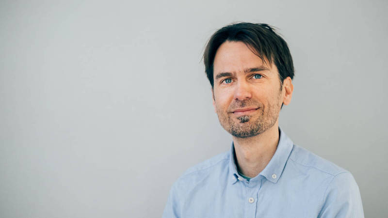
Portrait Alf Honigmann

© TUD CMCB, M. Gonciarz

We aim to understand how tissue architecture emerges from the structure and dynamics of cellular interfaces. My group develops a combination of super-resolution microscopy, organotypic tissue culture, genetics and biochemistry to understand the molecular underpinning of how cells control the physical properties of their cell membranes (lipid composition, adhesion, cortical tension and osmotic gradients) to shape tissues.

Our recent work on epithelial tight junctions revealed a new mechanism how adhesion complexes self-assemble via cell-cell contact mediated phase separation of scaffolding proteins. In addition, we have reconstructed the supra-molecular architecture of the apical junctional complex in 3D epithelial tissue, which provides an important molecular/structural resource to understand how epithelial cells couple cell-cell interfaces to the cytoskeleton, apical polarity and transcription. An exciting discovery is that we can control 3D tissue shapes by manipulating the molecular structure of tight junctions. We are now using this feature in a VW funded project to study and engineer tissue folding mechanisms in 3D tissue culture systems.

Overall, our approach to making scientific discoveries often involves the concept of reconstituting emergent functions from the interactions of its underlying components. The beauty of reconstitution experiments is that the overwhelming complexity of biological systems can be picked apart and broken down to essential mechanisms. We use this approach to understand how molecular interactions drive formation of supra-molecular complexes like adhesion junctions as well as how cellular interactions lead to organ specific tissue architectures.

Alf Honigmann Research: Figure 1
Figure 1: Research focus of the Honigmann lab. Most human organs are constructed from epithelial tissues that shape the internal body cavities. Organotypic (stem) cell culture allows us to reconstitute key features of organ formation under controlled conditions in cell culture. We use super-resolution microscopy to quantify the molecular organization of epithelial cell membranes during the process of tissue formation. From this we generate structural models of the mesoscale organization of adhesion complexes with molecular resolution. Using genetic perturbations and biochemical reconstitutions of key components we ask: What are the underlying mechanisms that drive assembly of mesoscale structures at membranes and how do the tissue shaping forces arise from and feedback to the molecular level?
Alf Honigmann Research: Figure 2 Dynamics of the scaffold protein ZO1 during tight junction formation in epithelial monolayer. ZO1 forms liquid-like condensates at cell-cell contact regions which elongate and fuse around the cell perimeters to form a tight junctional belt (see Beutel et al. 2019).
Alf Honigmann Research: Figure 3 First 2 days of pluripotent mouse embryonic stem cells in organoid culture forming the epiblast cavity. Cells were imaged using SPIM and cell interfaces were segmented using custom software.

Future Projects and Goals

  • Role of membrane remodelling in polarization and lumen formation in organoids
  • Understanding how cells measure and control physical properties of cell their interfaces during morphogenesis (organoids).  
  • Visualizing lipid trafficking and metabolism in cells and tissues with high resolution microscopy

Methodological and Technical Expertise

  • 3D organoid culture and genetic engineering
  • Microscopy: STED, SPIM, FLIM, FCS…
  • Laser ablation, photo-conversion, opto-genetics
  • Protein purification and reconstitution with membranes
  • Image analysis

CV

Since 2022
Professor of Biophysics at BIOTEC Dresden

2015–2022
Group leader at the Max-Planck-Institute of Molecular Cell Biology and Genetics in Dresden

2011–2015
Post-doctoral Fellow at the Max-Planck-Institute Biophysical-Chemistry with Stefan Hell, Goettingen

2010
PhD in Cell Biology and Biophysics, University of Osnabrueck

More Information

tu-dresden.de

Selected Publications

Beutel O, Maraspini R, Pombo-García K, Martin-Lemaitre C, Honigmann A
Phase Separation of Zonula Occludens Proteins Drives Formation of Tight Junctions
Cell. 179 (2019)

Sun D, LuValle-Burke I, Pombo-García K, Honigmann A
Biomolecular condensates in epithelial junctions
Current Opinion in Cell Biology 77, 102089 (2022)

Maraspini R, Wang CH, Honigmann A
Optimization of 2D and 3D cell culture to study membrane organization with STED microscopy
J. Phys. D. Appl. Phys. 53 (2019)

Gao M, Maraspini R, Beutel O, Zehtabian A, Eickholt B, Honigmann A, Ewers H
Expansion Stimulated Emission Depletion Microscopy (ExSTED)
ACS Nano. 12 (2018)

Honigmann A, Sadeghi S, Keller J, Hell SW, Eggeling C, Vink R
A lipid bound actin meshwork organizes liquid phase separation in model membranes
Elife. 3 (2014)

Contact

Biotechnology Center of the TU Dresden (BIOTEC)
Tatzberg 47/49
01307 Dresden